Since becoming a science-illustration professor my focus has shifted towards nature and biological science subjects, and my multi-faceted role as a consultant for an exhibitry-design company. However medical illustration has played a huge role in making me who & what I am, so I’m including in this gallery a few longtime favourites from my days in the Master of Science in Biomedical Communications (MScBMC) program at the University of Toronto.
I also have an enduring love for life drawing, particularly anatomical. In the context of illustration, life drawings are usually considered exercises rather than finished works. But if you think of the visual arts in similar terms to other arts such as music or dance, technique exercises are a fundamental part of the art form and contain a beauty all their own. I also find life drawing an intensely meditative experience. By that I don’t mean a mind-emptying experience; quite the contrary, it’s an experience that seems to make the conscious complications of life disappear in a concentrated focus on form, line and light.
Click on any image to launch full size slideshow. When finished, close slideshow by clicking on the x in the upper R corner.
All images ©Kathryn Chorney.
-
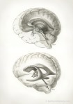
- Conceptual view of the fluid-filled ventricles of the brain. In textbooks they are usually shown as though they were floating structures, similar to how I’ve shown them in the lower figure. However I was more interested in showing how these shapes are formed by the structures that surround them. Carbon dust and gouache on strathmore plate finish bristol.
-
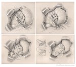
- Four of six figures from a surgical illustration series on kidney transplantation. Carbon pencil, carbon dust, and highlight on Canson paper.
-

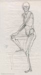
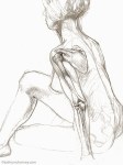
- Seated ‘skellyhead’, 10-minute study. I like to sketch in the skeletal elements from memory while drawing the live model. Conte on newsprint.
-
- A 10-minute study. I like to sketch in the skeletal elements from memory while drawing the live model. Conte on newsprint.
-

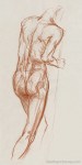
- Another 10-minute study, with skeletal elements sketched in from memory while observing the live model. Conte on newsprint.
-

- Anatomical drawing – male lower limbs. Ballpoint ink on sketchbook paper.
-
- Life study, with skeletal anatomy added later on tracing-paper overlay, then scanned and composited. Graphite on paper.
-
- Life study, with skeletal anatomy added later on tracing-paper overlay, then scanned and composited. Graphite on paper.
-
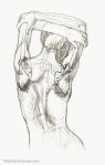
- Study of the surface anatomy of the back, drawn at anatomical life drawing workshop. Conte on newsprint.
-

- A 5-minute anatomical life study, with skeletal elements sketched in from memory while observing the live model. Conte on newsprint.
